Microscopic Techniques for Observing Cell Color

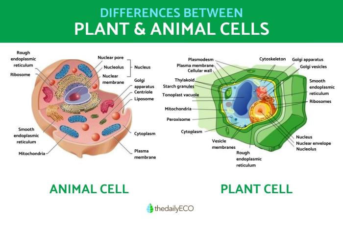
Coloring the cell: animal and plant – The vibrant tapestry of life, revealed at the cellular level, unveils a breathtaking spectrum of colors. These colors, arising from pigments within cells, hold vital clues to cellular function and health. To unravel these secrets, we turn to the powerful tools of microscopy, each offering a unique perspective on the colorful world within. Let us delve into the spiritual journey of understanding these microscopic techniques, recognizing the divine artistry present in even the smallest of life’s creations.
Just as a painter uses different brushes and paints to create a masterpiece, scientists employ various microscopy techniques to reveal the intricate details of cellular coloration. Each method illuminates different aspects of the cell’s pigmented landscape, allowing us to appreciate the divine complexity of creation.
Bright-Field Microscopy
Bright-field microscopy is the most basic and widely accessible technique. Light passes directly through the specimen, and colored components absorb specific wavelengths, revealing their presence against a bright background. This simple yet elegant method is invaluable for observing the overall distribution of pigments within cells, providing a foundational understanding of their spatial arrangement. The simplicity of this technique makes it an excellent starting point for cellular observation, much like a beginner artist learning the fundamentals of color mixing.
Dark-Field Microscopy
In contrast to bright-field microscopy, dark-field microscopy enhances the contrast of transparent specimens by illuminating them obliquely. Only scattered light from the specimen enters the objective lens, resulting in a bright specimen against a dark background. This technique is particularly useful for visualizing fine details of pigmented structures within cells, such as the intricate networks of chromoplasts in plant cells, showcasing the exquisite detail of God’s design.
The enhanced contrast reveals subtleties often missed in bright-field microscopy, much like a master artist using subtle shading to bring depth to their work.
Fluorescence Microscopy
Fluorescence microscopy employs fluorescent dyes or proteins that emit light at specific wavelengths when excited by a light source. This technique allows for the highly specific visualization of particular pigments or cellular components. For example, specific fluorescent probes can be used to target and illuminate chlorophyll in plant cells, or specific cellular organelles involved in pigment production, revealing the precise location and interaction of these components within the complex cellular machinery.
This method is analogous to a skilled artisan using specialized tools to highlight specific aspects of their creation, revealing the intricate beauty of God’s handiwork.
Comparison of Microscopic Techniques, Coloring the cell: animal and plant
Each microscopic technique possesses unique strengths and limitations, making it suitable for specific applications. The choice of technique depends on factors such as the resolution required, the cost of equipment, and the type of cells being studied. Consider these tools as different instruments in an artist’s palette, each contributing to a complete understanding of the cellular masterpiece.
| Technique | Resolution | Cost | Applicability |
|---|---|---|---|
| Bright-field | Moderate | Low | Wide range of cell types |
| Dark-field | Moderate | Moderate | Transparent specimens, highlighting fine details |
| Fluorescence | High | High | Specific pigment visualization, localization of cellular components |
Applications of Cell Coloration Studies: Coloring The Cell: Animal And Plant

The vibrant hues revealed through cell coloration techniques are not merely aesthetically pleasing; they are windows into the intricate workings of life itself. Understanding these colors allows us to delve into the mysteries of health, disease, evolution, and the environment, revealing insights that can guide us towards a healthier and more sustainable future. This journey of discovery, much like a spiritual quest, requires patience, precision, and a deep respect for the delicate balance of life.
The applications of cell coloration extend far beyond the microscope, impacting numerous fields and enriching our understanding of the natural world. It is a testament to the power of observation and the beauty of scientific inquiry. Just as a skilled artist uses color to evoke emotion and convey meaning, so too do scientists utilize cell coloration to unlock the secrets held within the microscopic realm.
Cell Coloration in Medical Diagnosis
Cell coloration plays a crucial role in diagnosing various diseases. For instance, the Pap smear, a common screening test for cervical cancer, relies on staining techniques to identify abnormal cells. The distinctive coloration patterns of cancerous cells, differing from healthy cells, allow for early detection and timely intervention. Similarly, blood smears stained with Wright’s stain allow for the identification of different blood cell types, aiding in the diagnosis of blood disorders like leukemia.
The unique staining properties of specific cellular components, like the nucleus or mitochondria, provide invaluable diagnostic information. Consider the image of a stained tissue sample under a microscope: the stark contrast between the deep purple nuclei of healthy cells and the pale, irregularly shaped nuclei of cancerous cells speaks volumes. This visual difference, achieved through careful coloration, is a life-saving tool.
Tracking Cell Movement and Development
Imagine a time-lapse film of a developing embryo, each cell meticulously colored to follow its journey. This is the power of cell tracking through coloration. Fluorescent dyes, attached to specific proteins within cells, allow scientists to visualize cell movement and migration during development. This technique is vital for understanding processes like wound healing, where cells move to repair damaged tissue, and embryonic development, where cells differentiate into various tissues and organs.
The vibrant trails of fluorescently labeled cells provide a captivating visual record of dynamic cellular processes, offering invaluable insights into the choreography of life. For example, researchers studying neuronal development might use fluorescent dyes to track the growth of axons as they extend towards their target cells, illustrating the complex pathways that form the nervous system.
Cell Coloration in Agriculture and Environmental Science
The application of cell coloration extends beyond medicine, impacting agriculture and environmental science. In agriculture, staining techniques are used to assess the health of plants, identifying pathogens or nutrient deficiencies at the cellular level. This allows for early intervention, preventing widespread crop damage. In environmental science, cell coloration is used to study the effects of pollutants on aquatic organisms.
By examining the cellular changes in response to toxins, scientists can assess the level of environmental contamination and develop strategies for remediation. Imagine microscopic algae cells, their vibrant green replaced by a sickly yellow hue under the influence of a pollutant. This stark visual change, revealed through coloration, serves as a critical warning signal for environmental health.
Cell Coloration and Evolutionary Processes
The study of cell coloration provides valuable insights into evolutionary processes. By comparing the coloration patterns of cells across different species, researchers can trace evolutionary relationships and understand how cellular structures have evolved over time. For example, the comparison of chloroplast structure and coloration in different plant species can reveal evolutionary relationships and adaptations to diverse environments. The subtle differences in coloration, reflecting variations in cellular components and metabolic pathways, offer clues to the intricate history of life on Earth.
These microscopic variations, when viewed collectively, paint a larger picture of the evolutionary tapestry.
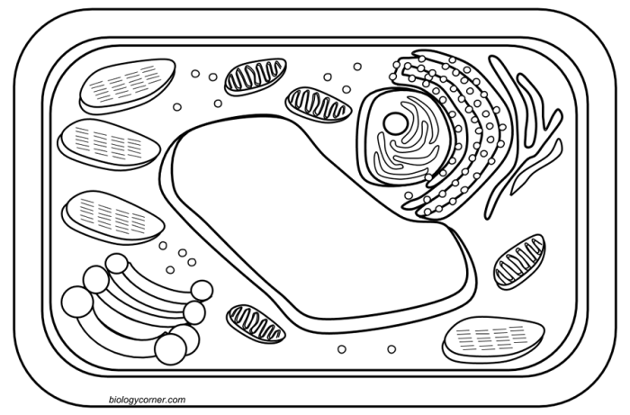
Understanding the vibrant hues of cells, whether in the intricate structures of plants or the fascinating complexity of animals, opens a world of wonder. To appreciate the diverse pigmentation of animal cells, consider the unique coloring of the axolotl, a creature whose beauty is readily explored through a delightful activity like the animal axolotl coloring page. This simple act of coloring helps visualize the distribution of pigments within a cell, bridging the gap between microscopic observation and artistic expression, further illuminating the beauty of cellular structures in both plant and animal life.


