Interpreting Color Coding Schemes

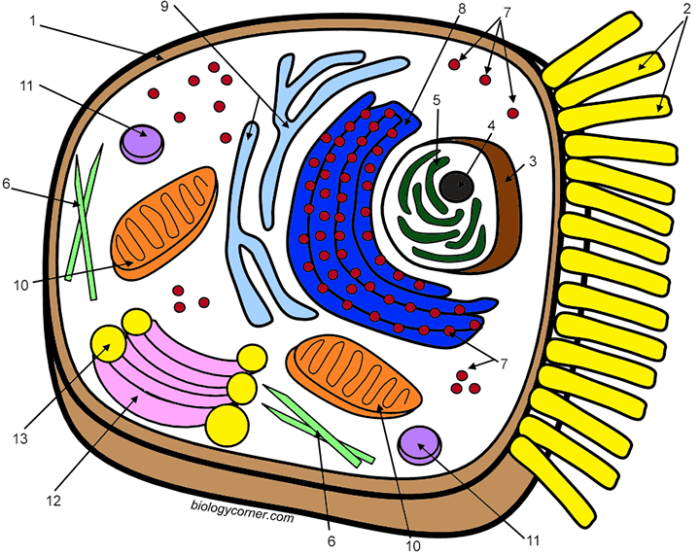
Animal cell coloring key page 30 – Effective color-coding in diagrams significantly impacts understanding, particularly for complex structures like animal cells. Choosing the right color scheme can illuminate relationships between organelles, highlight key features, and improve overall comprehension. Conversely, poor color choices can lead to confusion and misinterpretations. This section explores various color-coding strategies for animal cell diagrams and analyzes their advantages and disadvantages.Color schemes for animal cell diagrams should be carefully considered to enhance learning and understanding.
If you’re working on the animal cell coloring key, page 30 might present some challenges. Finding the correct identifications for each organelle can be tricky, but thankfully, there are resources available to help. For instance, you can check the comprehensive answer key provided by Biology Corner; a helpful resource is the animal cell coloring biology corner answer key.
Using this alongside your page 30 key should make completing your assignment much easier. Remember to double-check your work against both resources for accuracy.
Different approaches emphasize various aspects of the cell’s structure and function, leading to varied educational outcomes.
Color-Coding Schemes Based on Organelle Function
This approach uses colors to represent the primary function of each organelle. For instance, all organelles involved in energy production (mitochondria) could be shaded in a vibrant yellow, signifying energy and activity. Organelles responsible for protein synthesis (ribosomes, rough endoplasmic reticulum) might be depicted in a cool blue, representing the building blocks of life. The Golgi apparatus, involved in packaging and secretion, could be a warm orange, highlighting its role in cellular transport.
This scheme aids understanding by visually grouping organelles with similar roles, reinforcing functional relationships. A drawback might be that functionally similar organelles of varying sizes or locations may appear visually similar, potentially obscuring subtle differences.
Color-Coding Schemes Based on Organelle Size, Animal cell coloring key page 30
Here, color intensity or hue directly correlates with the relative size of organelles. Larger organelles, such as the nucleus, could be represented by a dark, saturated color, while smaller organelles, such as ribosomes, could be represented by a lighter, pastel shade of the same color. This method allows for a quick visual assessment of the relative scale of different organelles.
However, it may not be ideal for conveying functional relationships or locations within the cell. It could also be difficult to accurately represent the full range of sizes in a visually appealing and easily interpretable manner.
Color-Coding Schemes Based on Organelle Location
This method uses color to indicate the general location of organelles within the cell. For example, organelles predominantly found in the cytoplasm could be one color, while those associated with the cell membrane could be another. Organelles within the nucleus could be a third color. This approach is useful for highlighting spatial relationships and understanding the overall cellular organization.
A potential limitation is that it might not effectively communicate functional relationships or the relative size of different organelles. The visual distinction might also become less effective if many organelles occupy similar locations.
Benefits and Drawbacks of Different Color Schemes in Educational Materials
Using different color schemes offers several benefits. Visual learners benefit greatly from the use of color to categorize and organize information. Color can make complex structures more accessible and memorable. However, an improperly chosen color scheme can lead to confusion. For example, using colors that are too similar or that have strong emotional associations (e.g., using red for a benign structure) can hinder understanding.
Accessibility is also a crucial consideration. Colorblind individuals may not be able to distinguish between certain colors, making it vital to use colors with sufficient contrast and consider alternative methods of differentiation. Furthermore, the cultural context of color should be taken into account, as color symbolism varies across cultures.
Impact of Color Choice on Understanding Complex Biological Structures
Appropriate color choice is crucial for enhancing understanding of complex biological structures. A well-designed color scheme can highlight key features, relationships, and spatial arrangements, improving comprehension and retention. For example, using contrasting colors to differentiate between the nucleus and the cytoplasm can improve students’ ability to locate and identify these structures. Conversely, a poorly chosen color scheme can obscure important details and create confusion.
For instance, using similar colors for functionally distinct organelles can lead to misinterpretations of their roles. The careful selection of colors, considering factors such as contrast, accessibility, and cultural context, is therefore essential for creating effective and inclusive educational materials.
Illustrating an Animal Cell: Animal Cell Coloring Key Page 30

Animal cells, the fundamental building blocks of animal tissues and organs, are fascinatingly complex structures. They lack a rigid cell wall, unlike plant cells, giving them a more flexible and varied morphology. Understanding their internal organization is key to grasping their functions. This section will guide you through illustrating an animal cell, focusing on the accurate depiction of its organelles and their spatial relationships.
Animal cells are typically microscopic, ranging from 10 to 100 micrometers in diameter, though their size varies considerably depending on the cell type and its function. Their shape is also highly variable; some are spherical, others elongated, or even irregular, reflecting their specialized roles within the organism. Imagine a bustling miniature city, with various structures working in concert to maintain life.
This is analogous to the coordinated activity within an animal cell.
Animal Cell Organelle Arrangement and Appearance
The nucleus, the cell’s control center, is usually centrally located and appears as a large, round structure. It’s surrounded by a double membrane, the nuclear envelope, punctuated by nuclear pores that regulate the passage of molecules. The cytoplasm, a jelly-like substance filling the cell, contains a network of interconnected membranes called the endoplasmic reticulum (ER). The rough ER, studded with ribosomes (tiny granules responsible for protein synthesis), appears slightly darker and rougher than the smooth ER, which is involved in lipid metabolism and detoxification.
The Golgi apparatus, a stack of flattened sacs, resembles a series of interconnected pancakes and modifies and packages proteins. Mitochondria, the powerhouses of the cell, are sausage-shaped organelles with a folded inner membrane (cristae) that increase surface area for energy production. Lysosomes, small, membrane-bound sacs containing digestive enzymes, appear as tiny, spherical vesicles. Finally, the cytoskeleton, a network of protein filaments providing structural support and facilitating movement, is not easily visualized but is essential for maintaining cell shape and intracellular transport.
Step-by-Step Guide to Coloring an Animal Cell Diagram
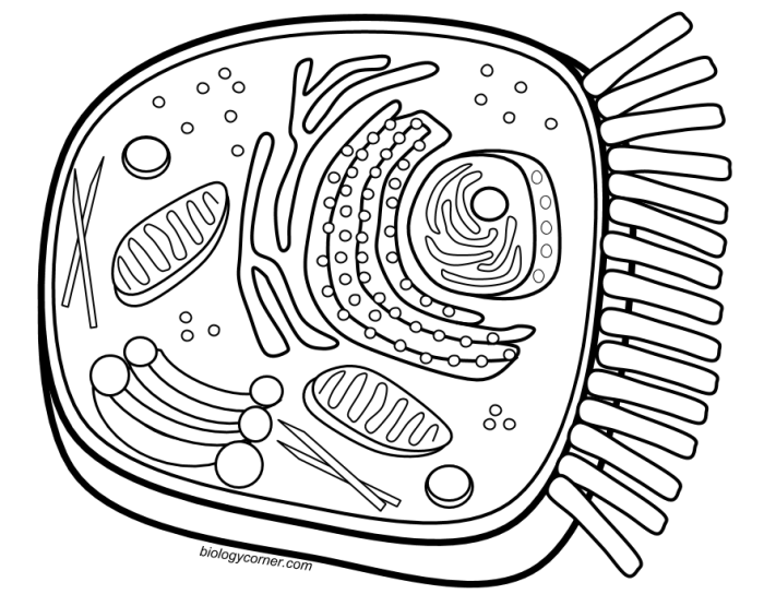
Assuming you have a pre-drawn diagram of an animal cell and a color key, follow these steps for accurate coloring:
- Identify Organelles: Carefully examine the diagram and identify each organelle (nucleus, mitochondria, Golgi apparatus, etc.).
- Consult the Color Key: Refer to your color key to determine the assigned color for each organelle. Ensure you understand the color coding scheme completely.
- Color Organelles: Using colored pencils, crayons, or markers, carefully color each organelle according to the color key. Try to maintain consistent coloring within each organelle.
- Label Organelles (Optional): If required, neatly label each organelle using a pencil or pen. Ensure labels are clear and concise.
- Review: Once completed, review your work to ensure all organelles are accurately colored and labeled according to the key.
Drawing an Animal Cell: A Step-by-Step Approach
Accurately drawing an animal cell requires attention to detail and a clear understanding of the relative sizes and locations of the organelles. The following steps will help guide you.
- Artikel the Cell Membrane: Begin by drawing a slightly irregular oval or circle to represent the cell membrane. Remember, animal cells lack a rigid cell wall, so the shape can be more flexible.
- Draw the Nucleus: Position a large, centrally located circle to represent the nucleus. Draw a double membrane around it, representing the nuclear envelope, and add a few small circles to depict nuclear pores.
- Add the Cytoplasm: Fill the space between the cell membrane and the nucleus with light shading to represent the cytoplasm.
- Illustrate the Endoplasmic Reticulum: Draw a network of interconnected membranes throughout the cytoplasm. Indicate the rough ER by adding small dots (ribosomes) along some of the membranes.
- Draw the Golgi Apparatus: Add a stack of flattened sacs near the nucleus, resembling a series of interconnected pancakes.
- Include Mitochondria: Draw several sausage-shaped organelles with folded inner membranes (cristae) scattered throughout the cytoplasm.
- Add Lysosomes: Include several small, spherical vesicles scattered throughout the cytoplasm, representing lysosomes.
FAQs
What is the best way to use this coloring key with younger students?
For younger students, simplify the key by focusing on only the major organelles. Use larger print and brighter colors. Consider incorporating a short, age-appropriate explanation of each organelle’s function.
Are there any online resources that offer similar animal cell coloring keys?
Yes, many educational websites and online resources provide printable animal cell diagrams and corresponding coloring keys. A simple web search should yield numerous results.
How can I assess student understanding after using the coloring key?
Assess understanding through quizzes, short answer questions, or by having students label a blank diagram of an animal cell. Observe their ability to correctly identify and describe the functions of the organelles.


