Introduction to Animal and Plant Cell Structures: Animal And Plant Cell Coloring Worksheet Pdf
Animal and plant cell coloring worksheet pdf – Cells are the fundamental building blocks of all living organisms. While all cells share some common features, there are significant differences between animal and plant cells, reflecting their distinct functions and lifestyles. Understanding these differences is crucial to comprehending the diversity of life.Animal and plant cells are both eukaryotic cells, meaning they possess a membrane-bound nucleus containing their genetic material.
However, their structures differ in several key aspects, primarily due to the differing needs of these two cell types. Plant cells need to support themselves against gravity and perform photosynthesis, leading to structural adaptations not found in animal cells.
Animal Cell Structures
Animal cells are characterized by their flexible cell membrane, a lack of rigid cell wall, and the presence of various organelles performing specific functions. Key components include the nucleus, which houses the DNA; mitochondria, the powerhouses generating energy; ribosomes, responsible for protein synthesis; the endoplasmic reticulum, involved in protein and lipid processing; and the Golgi apparatus, modifying and packaging proteins for secretion.
Lysosomes, containing digestive enzymes, break down waste materials, and the cytoskeleton provides structural support and facilitates movement within the cell.
Plant Cell Structures
Plant cells share many similarities with animal cells, but also possess unique features enabling them to carry out photosynthesis and provide structural support. Like animal cells, they have a nucleus, mitochondria, ribosomes, endoplasmic reticulum, and Golgi apparatus. However, they also contain chloroplasts, the sites of photosynthesis, where sunlight is converted into chemical energy. A large central vacuole regulates water balance and stores nutrients, and a rigid cell wall made primarily of cellulose provides structural support and protection.
Understanding the differences between animal and plant cell structures is often aided by visual learning tools like coloring worksheets. These PDFs provide a hands-on approach to grasping complex biological concepts. For a fun, related activity after completing your animal and plant cell worksheet, you might enjoy checking out these animal alphabet i coloring pages printable , offering a change of pace while still engaging with the animal kingdom.
Returning to the cellular level, remember that detailed coloring helps reinforce knowledge retention of the animal and plant cell structures.
Comparison of Animal and Plant Cells
The most striking differences between animal and plant cells lie in the presence or absence of certain key structures. Plant cells possess a cell wall, chloroplasts, and a large central vacuole, while animal cells lack these features. Animal cells, on the other hand, contain centrioles, which play a role in cell division, while plant cells generally do not. These structural differences reflect the distinct physiological needs and functions of each cell type.
Summary Table of Differences
| Feature | Animal Cell | Plant Cell |
|---|---|---|
| Cell Wall | Absent | Present (cellulose) |
| Chloroplasts | Absent | Present |
| Vacuoles | Small, numerous | Large, central |
| Centrioles | Present | Absent (usually) |
Worksheet Design and Functionality
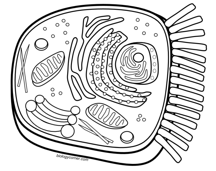
This section details the design and intended functionality of a coloring worksheet designed to enhance understanding of animal and plant cell structures. The worksheet employs a visual approach, combining engaging color schemes with clear labeling to facilitate learning across different age groups.The worksheet is designed to be a visually appealing and effective educational tool. It achieves this through a combination of strategic color choices, appropriate sizing of cellular components, and a logical layout that promotes comprehension.
Visual Elements and Design Choices
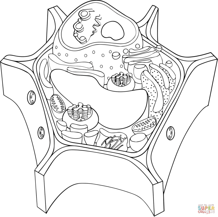
The worksheet features two large, side-by-side illustrations: one representing a typical animal cell and the other a typical plant cell. Each cell is depicted in a simplified, yet accurate, manner. The animal cell is primarily rendered in shades of light blue and purple, while the plant cell uses greens and browns to reflect its natural coloration. This color differentiation immediately distinguishes the two cell types.
Organelles are clearly Artikeld and oversized relative to their actual proportions, making them easily identifiable. Labels for major organelles, such as the nucleus, mitochondria, chloroplasts (in the plant cell), cell wall (plant cell), and vacuole (plant cell), are clearly printed near their corresponding structures, using a sans-serif font for easy readability. The layout is straightforward, with sufficient white space to prevent visual clutter.
The overall size of the worksheet is designed to be easily manageable for younger children, while still providing enough detail for older students.
Educational Use and Benefits
This coloring worksheet serves as an active learning tool, engaging students in a hands-on approach to understanding cell structures. The act of coloring reinforces visual memory and aids in associating labels with the specific organelles. The clear labeling and simplified illustrations ensure that even younger learners can grasp the fundamental differences between animal and plant cells. Older students can use the worksheet as a review tool, consolidating their knowledge of cell biology.
The visual nature of the worksheet also makes it accessible to visual learners.
Age Group Benefits
Young children (ages 5-8) can benefit from the simple illustrations and the act of coloring, helping them learn basic cell components and differentiate between animal and plant cells. Older children (ages 9-12) can utilize the worksheet to build upon their existing knowledge, focusing on the specific functions of organelles and comparing and contrasting animal and plant cells. Teenagers and adults can use the worksheet as a quick reference or review tool to refresh their understanding of fundamental cell biology.
The worksheet’s adaptability makes it suitable for a broad range of educational contexts, from homeschooling to classroom settings.
Educational Applications and Activities
This coloring worksheet offers a versatile tool for teaching about animal and plant cells across various grade levels. Its adaptability allows for differentiated instruction, catering to diverse learning styles and paces. The visual nature of the worksheet aids comprehension, particularly for younger learners, while the labeling activity encourages active learning and knowledge retention.The worksheet’s integration into a lesson plan can significantly enhance student understanding of cell structures and functions.
It can be used as a pre-activity to gauge prior knowledge, a main activity to reinforce learning, or a post-activity to assess comprehension. The flexibility of the worksheet allows for its seamless incorporation into various teaching methodologies.
Lesson Plan Integration Across Grade Levels
The worksheet can be effectively incorporated into lesson plans for different grade levels, adjusting the complexity and expectations accordingly. For elementary school (grades 3-5), the focus could be on identifying and coloring the major organelles, like the nucleus and cell wall (for plant cells). Middle school (grades 6-8) could include more detailed labeling of organelles and a comparison of animal and plant cells.
High school (grades 9-12) could incorporate advanced concepts, such as the functions of each organelle and the relationship between cell structure and function. For example, a high school lesson could include a discussion of how the structure of the mitochondria facilitates cellular respiration.
Classroom Activities Using the Worksheet
A series of classroom activities can build upon the worksheet to further reinforce learning. A simple activity involves students coloring the worksheet individually, then discussing their observations as a class. More engaging activities could include a collaborative labeling exercise, where students work in groups to identify and label the organelles, or a comparative analysis activity, where students compare and contrast animal and plant cells based on their completed worksheets.
A creative extension could involve students drawing their own cell diagrams, adding extra details learned from research.
Extension Activities for Early Finishers
Students who complete the worksheet quickly can be provided with extension activities to challenge and engage them further. These could include research projects on specific organelles, creating a three-dimensional model of a cell, or designing a presentation explaining the functions of different cell parts. Another possibility is researching diseases that affect cell function and presenting their findings to the class.
These activities cater to diverse learning styles and promote deeper understanding.
Assessing Student Understanding Using the Worksheet
The completed worksheet provides a valuable assessment tool. The accuracy of the coloring and labeling demonstrates the student’s understanding of cell structures and their locations. The teacher can assess student understanding by reviewing the completed worksheets, identifying areas where students may need further instruction or clarification. The worksheet also serves as a useful visual aid during discussions, allowing for targeted review of specific organelles and their functions.
This assessment method is both efficient and informative, offering direct insight into individual student learning.
Illustrative Examples
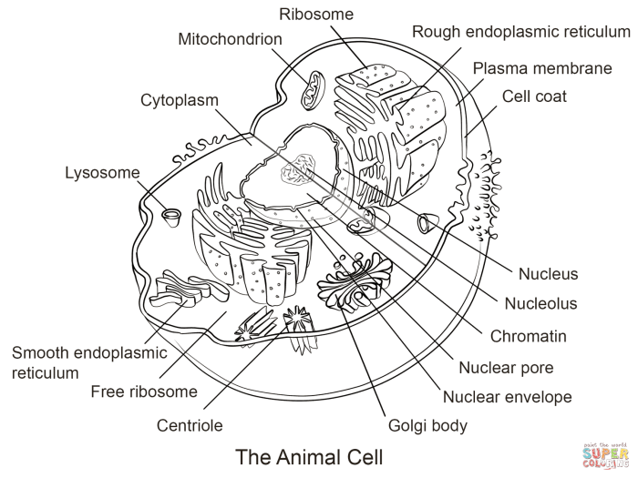
This section provides detailed descriptions of animal and plant cells, illustrating key organelles and their functions. We will also visually represent the processes of photosynthesis and cellular respiration. These descriptions aim to complement the coloring worksheet and enhance understanding of cell structure and function.
Labeled Diagram of an Animal Cell, Animal and plant cell coloring worksheet pdf
Imagine a microscopic, irregularly shaped blob, approximately 10-30 micrometers in diameter. This is a typical animal cell. At its center, you would find the nucleus, a relatively large, round organelle (5-10 micrometers) containing the cell’s genetic material (DNA). Surrounding the nucleus is the cytoplasm, a jelly-like substance filling the cell’s interior. Scattered throughout the cytoplasm are various organelles.
The mitochondria, often described as the “powerhouses” of the cell, are elongated, bean-shaped structures (0.5-10 micrometers) responsible for cellular respiration, generating energy (ATP). The endoplasmic reticulum (ER), a network of interconnected membranes, appears as a series of interconnected tubes and sacs. Rough ER, studded with ribosomes (tiny protein synthesis factories), is involved in protein synthesis and modification, while smooth ER plays a role in lipid metabolism and detoxification.
The Golgi apparatus, a stack of flattened sacs, modifies, sorts, and packages proteins for secretion or transport within the cell. Lysosomes, small, membrane-bound vesicles, contain digestive enzymes that break down waste materials and cellular debris. Finally, the cell membrane, a thin outer boundary, regulates the passage of substances into and out of the cell.
Labeled Diagram of a Plant Cell
A plant cell, typically rectangular or cube-shaped, is generally larger than an animal cell (10-100 micrometers). Like the animal cell, it possesses a nucleus, cytoplasm, mitochondria, endoplasmic reticulum, Golgi apparatus, and ribosomes, all performing similar functions. However, plant cells have unique features. Most prominently, a large central vacuole (20-80% of cell volume) occupies a significant portion of the cell’s interior, storing water, nutrients, and waste products.
The vacuole also contributes to cell turgor pressure, maintaining cell shape and rigidity. Surrounding the cell membrane is a rigid cell wall, composed primarily of cellulose, providing structural support and protection. Finally, chloroplasts, oval-shaped organelles (5-10 micrometers) containing chlorophyll, are responsible for photosynthesis. They are typically found near the periphery of the cell.
Visual Representation of Photosynthesis
Imagine a chloroplast, a green oval shape. Within this chloroplast, sunlight (represented by yellow arrows) enters and strikes chlorophyll molecules (represented by green dots). Water (H₂O, represented by blue circles) is absorbed through the roots and transported to the chloroplast. Carbon dioxide (CO₂, represented by red circles) enters the chloroplast through tiny pores on the leaf’s surface.
Through a complex series of chemical reactions (represented by interconnected lines and arrows), the energy from sunlight is used to convert water and carbon dioxide into glucose (C₆H₁₂O₆, represented by a six-sided sugar molecule) and oxygen (O₂, represented by two red circles bonded together). The glucose serves as food for the plant, and oxygen is released as a byproduct.
This entire process can be summarized as: 6CO₂ + 6H₂O + Light Energy → C₆H₁₂O₆ + 6O₂
Visual Representation of Cellular Respiration
Envision a mitochondrion, a bean-shaped organelle. Glucose (C₆H₁₂O₆, represented by a six-sided sugar molecule) enters the mitochondrion. Oxygen (O₂, represented by two red circles bonded together) diffuses into the mitochondrion from the surrounding cytoplasm. Through a series of chemical reactions (represented by interconnected lines and arrows), glucose is broken down, releasing energy in the form of ATP (represented by small yellow stars), water (H₂O, represented by blue circles), and carbon dioxide (CO₂, represented by red circles).
The ATP provides energy for various cellular processes. This process can be summarized as: C₆H₁₂O₆ + 6O₂ → 6CO₂ + 6H₂O + ATP (energy)
FAQ Corner
Can this worksheet be used for younger children?
Yes, the worksheet can be adapted for younger children by simplifying the labels and focusing on the major organelles. Adult supervision may be helpful.
Is the PDF printable?
Yes, the worksheet is designed for easy printing. Standard printer settings should suffice.
Are the answers provided?
While the worksheet itself doesn’t include answers, a separate answer key could be easily created by the educator for assessment purposes.
What software is needed to open the PDF?
Any PDF reader, such as Adobe Acrobat Reader (free), will work.

