Cilia Coloring Techniques in Microscopy

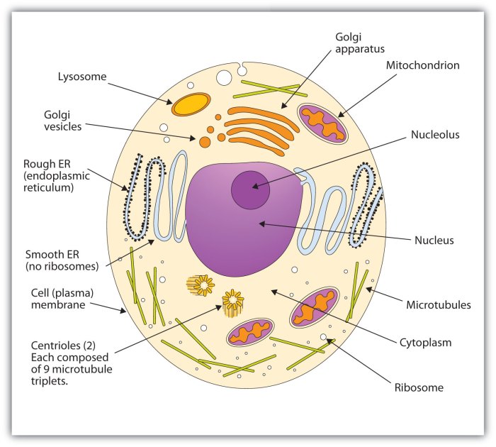
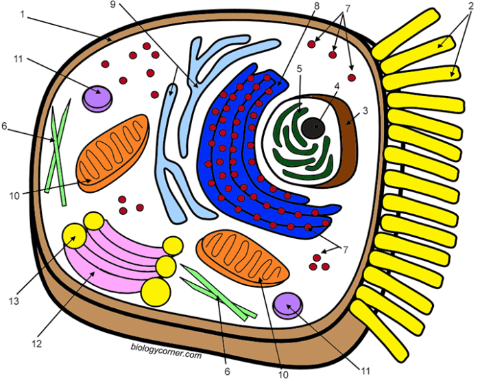
Animal cell coloring cilia – Visualizing cilia, the hair-like projections on the surface of many eukaryotic cells, requires specialized staining techniques in microscopy. These techniques enhance contrast and allow for detailed observation of cilia structure, arrangement, and function. The choice of staining method depends on the specific research question and the desired level of detail.
Staining Methods for Cilia Visualization
Several staining methods effectively highlight cilia in microscopy. These methods differ in their mechanisms of action, specificity, and the level of detail they provide. Commonly used stains include immunofluorescence, which uses antibodies to target specific cilia proteins, making them fluorescent; silver staining, a classic method that stains cilia darkly against a lighter background; and various dyes such as India ink, which can highlight cilia by negative staining.
Each method offers advantages and disadvantages depending on the application.
Comparison of Staining Effectiveness
Immunofluorescence offers high specificity, allowing researchers to target particular cilia proteins and study their localization and dynamics. However, it requires specialized antibodies and equipment, and the process can be more time-consuming and expensive. Silver staining is a simpler and more cost-effective technique, providing good contrast for cilia visualization. However, it lacks the specificity of immunofluorescence and may not be suitable for all types of cilia.
Dyes like India ink provide a quick and easy method for visualizing cilia via negative staining, showing their overall structure and arrangement, but offer less detailed information about their fine structure. The choice of stain depends on the specific needs of the experiment and available resources.
Sample Preparation for Cilia Observation
Proper sample preparation is crucial for successful cilia visualization. This involves carefully fixing the cells to preserve their structure, permeabilizing the cell membrane to allow the stain to penetrate, and mounting the sample appropriately for microscopy. Fixation methods vary depending on the cell type and the chosen staining technique. Common fixatives include formaldehyde and glutaraldehyde. Permeabilization often involves the use of detergents such as Triton X-100.
Understanding the intricate structure of animal cell cilia through coloring can be a fun and educational activity. The delicate hair-like projections are fascinating to visualize, and this detailed approach can be compared to the simpler, yet equally engaging, task of coloring animal babies coloring pages , where the focus shifts to the adorable features of young animals. Returning to the cellular level, accurate depiction of cilia helps in grasping their vital role in cell function.
Careful attention to these steps is crucial for maintaining cilia integrity and preventing artifacts.
Step-by-Step Protocol: Staining Cilia in Tracheal Epithelial Cells
This protocol Artikels the staining of cilia in human tracheal epithelial cells using immunofluorescence with an antibody targeting acetylated tubulin, a major component of cilia microtubules.
| Step | Reagent | Time | Observation |
|---|---|---|---|
| 1. Cell Fixation | 4% Paraformaldehyde in PBS | 15 minutes at room temperature | Cells become less transparent |
| 2. Permeabilization | 0.1% Triton X-100 in PBS | 5 minutes at room temperature | Cells become more permeable |
| 3. Blocking | 10% Normal Goat Serum in PBS | 30 minutes at room temperature | Reduces non-specific antibody binding |
| 4. Primary Antibody Incubation | Anti-acetylated tubulin antibody (diluted according to manufacturer’s instructions) | Overnight at 4°C | N/A (microscopic observation required later) |
| 5. Secondary Antibody Incubation | Fluorescently conjugated secondary antibody (e.g., Alexa Fluor 488) | 1 hour at room temperature | N/A (microscopic observation required later) |
| 6. Mounting | Antifade mounting medium with DAPI (for nuclear staining) | Immediately after washing | Cells are ready for microscopic observation |
The Role of Cilia in Cellular Processes

Cilia, hair-like structures extending from the surface of many animal cells, play multifaceted roles in cellular function, significantly impacting cell motility, fluid transport, and cellular signaling. Their dysfunction has profound consequences for overall cellular health and organismal well-being.
Cilia and Cell Motility
Cilia are crucial for the movement of certain single-celled organisms and for the movement of fluids across the surface of tissues in multicellular organisms. In single-celled organisms like Paramecium, coordinated beating of numerous cilia propels the cell through its environment. This coordinated movement results from a complex interplay of microtubules, dynein motor proteins, and calcium signaling. In multicellular organisms, the movement is often less about overall cell locomotion and more about moving substances across a cell surface.
Cilia and Fluid Movement
The rhythmic beating of cilia facilitates the movement of fluids across epithelial surfaces throughout the body. For example, in the respiratory tract, cilia beat in a coordinated fashion to move mucus containing trapped particles—like dust and pathogens—out of the lungs. Similarly, in the fallopian tubes, cilia propel the ovum towards the uterus. The efficiency of this fluid transport depends on the precise arrangement and coordinated beating of numerous cilia, highlighting the importance of their structural integrity and functional coordination.
Disruption of this coordinated movement can lead to significant health problems, as discussed later.
Cilia and Cellular Signaling Pathways
Cilia act as sensory organelles, receiving and transducing signals from the extracellular environment. They play a vital role in several signaling pathways, including the Hedgehog, Wnt, and planar cell polarity (PCP) pathways. These pathways are crucial for embryonic development, tissue patterning, and cell differentiation. Cilia function as antennas, receiving external signals and initiating intracellular signaling cascades that regulate gene expression and cellular processes.
Defects in ciliary signaling can lead to developmental abnormalities and various diseases.
Consequences of Cilia Dysfunction
Dysfunctional cilia result in a range of diseases collectively known as ciliopathies. These disorders manifest in diverse ways depending on the specific ciliary component affected and the tissues involved. For instance, defects in ciliary structure or function can lead to respiratory infections due to impaired mucus clearance (primary ciliary dyskinesia), polycystic kidney disease (PKD) due to impaired fluid flow in the kidneys, and various developmental abnormalities, such as situs inversus (reversed organ placement).
The severity of ciliopathies varies greatly, ranging from mild to life-threatening conditions, underscoring the crucial role of cilia in maintaining cellular and organismal health.
Cilia and Disease

Cilia, those tiny hair-like structures projecting from the surface of many cells, play crucial roles in various physiological processes. Disruptions in their structure or function, however, can lead to a range of debilitating diseases, collectively known as ciliopathies. These conditions highlight the essential contribution of cilia to human health.Cilia defects manifest in diverse ways depending on the specific gene mutation and the type of cilia affected (motile or primary).
The severity of symptoms also varies considerably, ranging from mild to life-threatening. Understanding the relationship between specific cilia defects and the resulting diseases is critical for developing effective diagnostic tools and therapeutic strategies.
Cilia Defects and Specific Diseases
The connection between cilia dysfunction and disease is multifaceted. Mutations in genes encoding proteins involved in cilia structure, assembly, or function can cause a spectrum of disorders. For instance, defects in genes responsible for intraflagellar transport (IFT), a crucial process for cilia formation and maintenance, are linked to a wide array of ciliopathies. Similarly, mutations affecting the dynein arms, responsible for ciliary motility, can lead to specific motility-related disorders.
Symptoms Associated with Cilia-Related Disorders
Symptoms associated with ciliopathies are highly variable and depend on the affected organ system and the specific genetic defect. Some common features include respiratory problems (chronic bronchitis, sinusitis), impaired vision (retinitis pigmentosa), hearing loss, kidney abnormalities (polycystic kidney disease), and skeletal abnormalities. Furthermore, some ciliopathies present with multi-system involvement, affecting multiple organs simultaneously. For example, Bardet-Biedl syndrome, a severe ciliopathy, can manifest with retinal degeneration, obesity, polydactyly (extra fingers or toes), intellectual disability, and kidney disease.
In contrast, primary ciliary dyskinesia (PCD) primarily affects the respiratory system, leading to chronic respiratory infections and reduced lung function.
Current Treatments and Therapies for Diseases Linked to Cilia Dysfunction
Currently, there is no cure for most ciliopathies. Treatment strategies primarily focus on managing symptoms and improving the patient’s quality of life. This often involves multidisciplinary approaches, incorporating respiratory therapy, ophthalmological care, renal management, and genetic counseling. For respiratory issues associated with PCD, therapies may include bronchodilators, mucolytics, and antibiotics to combat recurrent infections. For kidney-related problems, dialysis or kidney transplantation may be necessary.
Research into gene therapy and other innovative approaches holds promise for future treatments, but these are still in early stages of development. In some cases, supportive care and management of complications are the mainstays of treatment.
List of Diseases Related to Cilia Dysfunction, Animal cell coloring cilia
The following list provides a sample of diseases associated with cilia dysfunction. This is not an exhaustive list, as many other conditions are linked to cilia defects.
- Primary Ciliary Dyskinesia (PCD): Characterized by impaired motility of cilia in the respiratory tract, leading to recurrent respiratory infections.
- Kartagener Syndrome: A form of PCD associated with situs inversus (reversed organ placement).
- Bardet-Biedl Syndrome (BBS): A multi-system disorder with retinal degeneration, obesity, polydactyly, intellectual disability, and kidney disease.
- Alstrom Syndrome: Another multi-system disorder affecting vision, hearing, and the heart, along with diabetes and obesity.
- Joubert Syndrome: A neurological disorder characterized by a distinctive midbrain-hindbrain malformation.
- Nephronophthisis: A cystic kidney disease that primarily affects the kidneys.
User Queries: Animal Cell Coloring Cilia
What is the typical size of a cilium?
Cilia are typically 5-10 micrometers in length and 0.25 micrometers in diameter.
What are some examples of diseases caused by cilia dysfunction?
Primary ciliary dyskinesia (PCD), Kartagener syndrome, and polycystic kidney disease are examples of diseases linked to cilia dysfunction.
Can cilia regenerate?
Yes, under certain conditions, cilia can regenerate, although the process is complex and depends on various factors.
What is the difference between cilia and flagella?
While both are hair-like appendages, cilia are typically shorter and more numerous than flagella, and they often beat in a coordinated manner.


