Introduction to Animal Cell Structure: Animal Cell Coloring Page Biology Corner
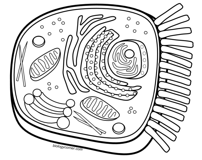
Animal cell coloring page biology corner – Animal cells are the fundamental building blocks of animals, exhibiting a complex internal organization crucial for their diverse functions. Understanding their structure is key to grasping the intricacies of animal biology. This section will explore the major organelles within an animal cell and their roles.
Animal cells, unlike plant cells, lack a cell wall and chloroplasts. Their structure is characterized by a flexible membrane enclosing a variety of specialized compartments, each performing specific tasks. The efficient coordination of these organelles ensures the cell’s survival and function.
Major Organelles of an Animal Cell
The following table details the key organelles found within a typical animal cell, their functions, and brief descriptions. Note that not all animal cells will contain every organelle listed, and the relative abundance of each can vary depending on the cell type and its function.
| Organelle | Function | Description |
|---|---|---|
| Nucleus | Houses genetic material (DNA); controls cell activities. | The control center of the cell, containing chromosomes and the nucleolus, which is involved in ribosome production. |
| Ribosomes | Protein synthesis. | Small structures found free in the cytoplasm or attached to the endoplasmic reticulum, responsible for translating genetic information into proteins. |
| Endoplasmic Reticulum (ER) | Protein and lipid synthesis; transport of molecules. | A network of membranes extending throughout the cytoplasm. Rough ER (with ribosomes) synthesizes proteins, while smooth ER synthesizes lipids and detoxifies substances. |
| Golgi Apparatus (Golgi Body) | Processes and packages proteins and lipids. | Modifies, sorts, and packages proteins and lipids for secretion or delivery to other organelles. Think of it as the cell’s “post office.” |
| Mitochondria | Cellular respiration; ATP production. | The “powerhouses” of the cell, generating energy (ATP) through cellular respiration. They have their own DNA. |
| Lysosomes | Waste breakdown; digestion of cellular components. | Membrane-bound sacs containing enzymes that break down waste products and cellular debris. |
| Cytoplasm | Supports organelles; site of many metabolic reactions. | The jelly-like substance filling the cell, containing the organelles and providing a medium for cellular processes. |
| Cell Membrane | Regulates what enters and leaves the cell. | A selectively permeable membrane surrounding the cell, controlling the passage of substances into and out of the cell. |
| Centrioles | Involved in cell division. | Paired cylindrical structures involved in organizing microtubules during cell division. |
Differences Between Plant and Animal Cells
While both plant and animal cells are eukaryotic (possessing a membrane-bound nucleus), they exhibit several key differences. These differences reflect the distinct needs and functions of these two cell types.
Plant cells possess a rigid cell wall made of cellulose, providing structural support and protection. This contrasts sharply with the flexible cell membrane of animal cells. Furthermore, plant cells contain chloroplasts, the organelles responsible for photosynthesis, allowing them to produce their own food. Animal cells lack chloroplasts and rely on consuming other organisms for energy. Plant cells typically have a large central vacuole for storing water and nutrients, a feature less prominent in animal cells.
The presence or absence of these organelles significantly influences the overall structure and function of plant and animal cells.
Designing a Coloring Page
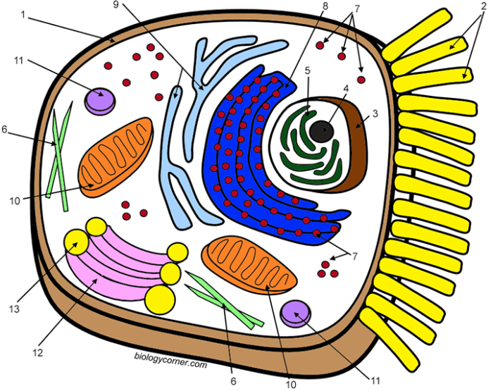
Creating an engaging and informative animal cell coloring page requires careful consideration of both accuracy and visual appeal. The design should accurately reflect the structure of an animal cell while remaining accessible and enjoyable for the intended audience. This involves creating both a detailed version for older learners and a simplified version for younger children.Designing a coloring page necessitates a thoughtful approach to visual representation, ensuring clarity and accuracy in depicting the cell’s organelles.
The choice of color and layout significantly impacts the overall effectiveness of the learning tool. The key is to balance educational rigor with visual appeal to maximize engagement.
Detailed Animal Cell Coloring Page
This version will include a comprehensive representation of the major organelles within an animal cell. The organelles should be clearly delineated and labeled, with a separate key provided for easy identification. The cell membrane should be depicted as a continuous outer boundary, enclosing all the organelles. The nucleus, a large, centrally located organelle, should be clearly shown. Other organelles such as the mitochondria (represented as bean-shaped structures), the endoplasmic reticulum (a network of interconnected membranes), the Golgi apparatus (a stack of flattened sacs), ribosomes (small dots), lysosomes (small circles), and the vacuoles (smaller than those found in plant cells) should be included and accurately positioned within the cell.
The key should list each organelle with its name and a brief description of its function. For example, the mitochondria could be described as the “powerhouses” of the cell. The overall design should be visually appealing, using a color scheme that is both attractive and conducive to learning. The cell’s cytoplasm should be filled with subtly shaded color to distinguish it from the organelles.
Exploring the intricacies of animal cell structure can be made engaging through the use of coloring pages. A readily available resource for such activities is the animal cell coloring page from Biology Corner, which provides a great starting point for learning. You can find a helpful version at animal cell coloring biologycorner com , offering a detailed illustration to color and label.
These coloring pages, in turn, serve as an effective tool for understanding the components and functions of an animal cell, enhancing comprehension of biology concepts.
Simplified Animal Cell Coloring Page, Animal cell coloring page biology corner
For younger learners, a simplified version is essential. This version will focus on only the most prominent organelles: the nucleus, the cell membrane, and the cytoplasm. The nucleus can be depicted as a large, centrally located circle, clearly differentiated from the cytoplasm. The cell membrane can be represented as a simple outer boundary. The cytoplasm can be represented as a shaded area surrounding the nucleus.
The key for this simplified version will only include these three major components. The overall design should be simple, clean, and engaging for young children, using bright, attractive colors. Avoiding excessive detail will make the coloring page less overwhelming and easier for children to understand.
Creating a High-Quality Printable Coloring Page
Creating a high-quality printable coloring page involves several key steps. First, the design should be created using a vector graphics program such as Adobe Illustrator or Inkscape. This ensures that the image can be scaled to any size without losing quality. The organelles should be clearly Artikeld and easily distinguishable from each other. The lines should be thick enough to be easily colored in by children, but not so thick that they overwhelm the design.
Next, a key should be created that clearly identifies each organelle. This key should be placed either within the coloring page itself or on a separate sheet. Finally, the coloring page should be saved as a high-resolution PDF file to ensure that it prints clearly and accurately. The color palette should be considered carefully; bright, non-clashing colors are preferable for younger children.
The file should be tested on various printers to ensure consistent print quality.
Educational Applications of the Coloring Page
This animal cell coloring page offers a versatile tool for educators to enhance student learning in various ways. Its engaging visual nature makes it particularly effective in reinforcing key concepts related to animal cell structure and function, catering to diverse learning styles and making complex biological information more accessible.The coloring page can be effectively integrated into a variety of teaching methodologies, stimulating active learning and improving knowledge retention.
It’s not just a passive activity; it encourages students to actively engage with the material, improving comprehension and recall.
Using the Coloring Page to Enhance Understanding
The act of coloring the different organelles of the animal cell requires students to repeatedly interact with their names and functions. By associating colors with specific structures (e.g., a vibrant green for the chloroplast, if applicable, or a bright blue for the nucleus), students create a visual mnemonic that aids memorization. This hands-on approach transforms a potentially abstract concept into a concrete, memorable experience.
Furthermore, the coloring page can be used as a springboard for discussions about the roles of each organelle within the cell, prompting students to explore the interconnectivity of cellular processes. For example, coloring the mitochondria could lead to a discussion about cellular respiration and energy production.
Incorporating the Coloring Page into Different Teaching Methodologies
The coloring page is adaptable to various teaching styles. In a direct instruction model, the coloring activity can serve as a reinforcement exercise after a lecture or presentation on animal cell structure. In a more inquiry-based approach, students can use the coloring page as a guide to research and label the organelles independently, promoting self-directed learning. Collaborative learning can be facilitated by having students work in pairs or small groups, discussing the functions of each organelle and coordinating their coloring efforts.
This collaborative effort encourages peer teaching and learning.
Assessing Student Learning with the Coloring Page
The coloring page itself can be a simple assessment tool. Teachers can observe whether students correctly identify and color the various organelles, providing a quick visual check of understanding. More formal assessment can be incorporated by adding a labeling component, where students write the names of the organelles next to their colored representations. This provides a more detailed assessment of their knowledge.
Alternatively, the coloring page can be used as a pre-assessment to gauge prior knowledge before introducing more complex concepts, or as a post-assessment to measure learning gains after a lesson. For instance, comparing the pre- and post-coloring page activities can reveal the effectiveness of the teaching strategies employed.
Questions and Answers
What age group is this coloring page suitable for?
The coloring page is adaptable for various age groups. A simplified version is provided for younger learners, while a more detailed version caters to older students.
Are there any printable versions available?
The intention is to create a high-quality, printable coloring page suitable for educational use. The design should be optimized for clear printing.
Can this coloring page be used for assessment?
Yes, the coloring page can be used as a formative assessment tool. Teachers can assess students’ understanding of organelles and their functions based on their coloring and labeling accuracy.

