Understanding Animal Cell Structure

Animal cell coloring pdf function answer key – Animal cells, the fundamental building blocks of animals, are complex structures containing a variety of organelles, each with a specific function contributing to the overall health and operation of the cell. Understanding these components is crucial to grasping the intricacies of life itself.
The cell membrane, also known as the plasma membrane, is a selectively permeable barrier that encloses the cell’s contents. It regulates the passage of substances into and out of the cell, maintaining a stable internal environment. This process, crucial for cell survival, is known as homeostasis. The membrane is composed of a phospholipid bilayer with embedded proteins that facilitate transport, communication, and other essential functions.
The selective permeability ensures that necessary nutrients enter and waste products exit the cell while maintaining a balance of ions and molecules inside.
Major Organelles and Their Functions
Animal cells possess numerous organelles, each performing a specific role. These can be grouped into functional categories for easier understanding.
Energy Production
The powerhouse of the cell, the mitochondrion, is responsible for generating adenosine triphosphate (ATP), the cell’s primary energy currency. Through cellular respiration, mitochondria break down glucose and other fuel molecules to produce ATP, which powers cellular processes.
Protein Synthesis
Protein synthesis involves two major organelles: the ribosomes and the endoplasmic reticulum (ER). Ribosomes, either free-floating in the cytoplasm or bound to the ER, synthesize proteins according to instructions from the cell’s DNA. The rough endoplasmic reticulum (RER), studded with ribosomes, modifies and transports newly synthesized proteins. The smooth endoplasmic reticulum (SER) synthesizes lipids and detoxifies harmful substances.
Genetic Material and Control
The nucleus houses the cell’s genetic material, DNA, organized into chromosomes. It controls gene expression, regulating the synthesis of proteins and other cellular activities. The nucleolus, a dense region within the nucleus, is responsible for ribosome production.
Waste Processing and Storage
Lysosomes are membrane-bound organelles containing digestive enzymes that break down waste materials, cellular debris, and foreign substances. The Golgi apparatus modifies, sorts, and packages proteins and lipids for secretion or transport to other organelles.
Structural Support and Movement
The cytoskeleton, a network of protein filaments, provides structural support and helps maintain cell shape. It also plays a role in cell movement and intracellular transport. Centrioles, found near the nucleus, are involved in cell division.
Cell Membrane and Homeostasis
The cell membrane is a fluid mosaic of phospholipids and proteins. Its selective permeability is crucial for maintaining homeostasis. This involves regulating the concentration of ions, water, and other molecules within the cell. Transport proteins embedded in the membrane facilitate the movement of specific substances across the membrane, either passively (e.g., diffusion, osmosis) or actively (e.g., active transport, requiring energy).
The membrane also plays a role in cell signaling, receiving and transmitting signals from the external environment.
Differences Between Plant and Animal Cells
While both plant and animal cells are eukaryotic, possessing membrane-bound organelles, they differ significantly in several key aspects. Plant cells have a rigid cell wall made of cellulose, providing structural support and protection. They also contain chloroplasts, which conduct photosynthesis, converting light energy into chemical energy. Large central vacuoles are characteristic of plant cells, storing water, nutrients, and waste products.
Animal cells lack these structures.
Finding the answers for your animal cell coloring pdf function worksheet can be challenging, but understanding the structures is key. A helpful resource for this is comparing and contrasting animal and plant cells, which you can explore further by checking out this excellent guide on animal and plant cell coloring and comparison. This comparison will solidify your understanding of the unique features of animal cells, ultimately making those answer keys much easier to decipher.
Therefore, mastering the animal cell coloring pdf function answer key becomes significantly more manageable.
Simple Diagram of an Animal Cell
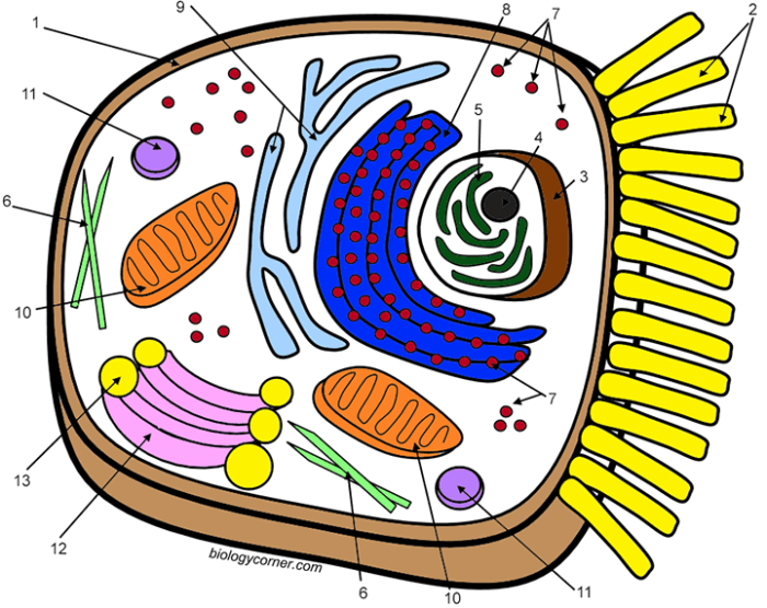
Imagine a circle representing the cell membrane. Within this circle, draw smaller circles and ovals of varying sizes to represent the organelles. Label the nucleus (a large, centrally located circle), the mitochondria (smaller, bean-shaped structures scattered throughout), the ribosomes (small dots, some attached to the endoplasmic reticulum – a network of interconnected membranes), the Golgi apparatus (a stack of flattened sacs), and the lysosomes (small, membrane-bound vesicles).
You can also include a representation of the cytoskeleton (a network of fine lines) and the centrioles (small cylindrical structures near the nucleus).
Coloring Activities and Their Educational Value

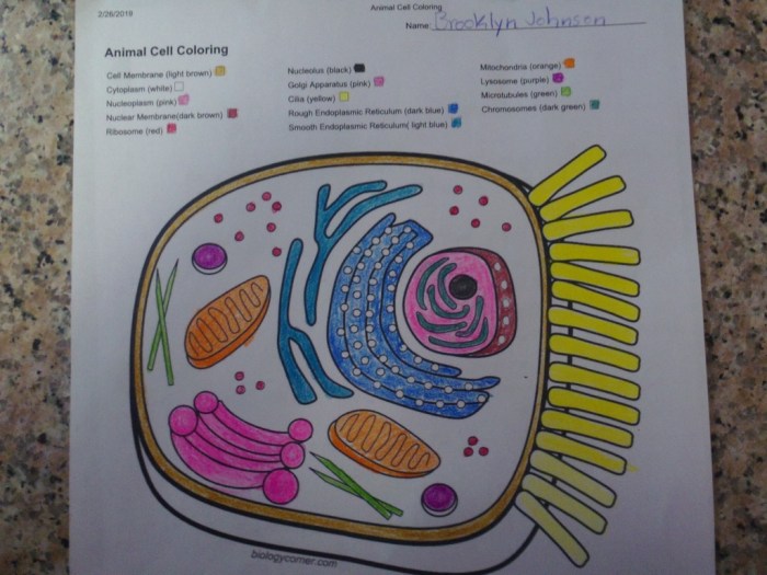
Coloring activities, often associated with childhood leisure, offer a surprisingly effective tool for enhancing learning, particularly in science education. The act of coloring engages multiple senses and cognitive processes, leading to improved understanding and retention of complex biological concepts like animal cell structure. This method transforms passive learning into an active, hands-on experience, making abstract ideas more concrete and accessible.Coloring activities significantly enhance understanding of animal cell structure by transforming abstract concepts into visually engaging representations.
Students actively participate in constructing their understanding as they color and label the different organelles, solidifying their knowledge through a kinesthetic learning experience. This process moves beyond simple memorization; it fosters a deeper understanding of the relationships between different parts of the cell and their functions.
Memory Retention of Biological Concepts Through Coloring
Coloring aids memory retention by engaging multiple cognitive pathways simultaneously. The visual act of coloring reinforces the visual representation of the cell structure, while the act of labeling activates verbal memory. This multi-sensory approach creates stronger neural connections, improving long-term retention. For instance, a student who colors a mitochondrion and labels it “powerhouse of the cell” is more likely to remember its function than a student who only reads about it.
The association between the visual image, the written label, and the function creates a robust memory trace.
Benefits of Hands-On Activities in Science Education
Hands-on activities like coloring provide invaluable benefits in science education. They cater to diverse learning styles, making complex concepts accessible to visual, kinesthetic, and tactile learners. The active engagement fosters a deeper understanding and greater enthusiasm for the subject matter compared to passive learning methods like simply reading a textbook. The process of coloring encourages exploration and experimentation, allowing students to discover and correct their own understanding.
This active involvement promotes critical thinking and problem-solving skills.
Comparison of Different Coloring Methods for Enhancing Learning
Different coloring methods can further enhance learning outcomes. For example, using different colors for different organelles can improve differentiation and memorization. Students could use a color-coding system, assigning specific colors to specific organelles consistently across all their work. Alternatively, incorporating shading techniques to represent three-dimensionality can create a more realistic and memorable representation of the cell structure.
Simple line drawings can be used for younger students, while more detailed diagrams can challenge older students. The choice of method should be tailored to the age and understanding of the students.
Learning Objectives Achievable Through Cell Coloring Exercises
The following learning objectives can be effectively achieved through cell coloring exercises:
- Identify and label major organelles of an animal cell (e.g., nucleus, mitochondria, ribosomes, endoplasmic reticulum, Golgi apparatus).
- Describe the function of each major organelle within the animal cell.
- Understand the relationship between the structure and function of each organelle.
- Compare and contrast the structures and functions of different organelles.
- Develop a deeper understanding of the overall organization and function of the animal cell.
Illustrating Key Organelles
Understanding the structure and function of key organelles is crucial for comprehending the complexities of animal cell biology. This section provides detailed descriptions of several vital organelles, emphasizing their roles in maintaining cellular processes. These descriptions will enhance understanding when used in conjunction with a cell diagram or coloring activity.
The Nucleus: Control Center of the Cell
The nucleus, often described as the cell’s “control center,” is a large, membrane-bound organelle that houses the cell’s genetic material, DNA. It’s typically spherical or oval in shape and is the most prominent feature within most eukaryotic cells. The nuclear envelope, a double membrane perforated by nuclear pores, regulates the passage of molecules between the nucleus and the cytoplasm.
Within the nucleus, DNA is organized into chromosomes, which are condensed structures of DNA and proteins. DNA replication, the process of creating an exact copy of the DNA molecule, occurs within the nucleus during the S phase of the cell cycle. This precise replication ensures that each daughter cell receives a complete set of genetic instructions. The nucleolus, a dense region within the nucleus, is the site of ribosome biogenesis – the production of ribosomes.
Mitochondria: Powerhouses of the Cell
Mitochondria are double-membrane-bound organelles often referred to as the “powerhouses” of the cell. Their inner membrane is highly folded into cristae, significantly increasing the surface area for cellular respiration. This process, which converts nutrients into adenosine triphosphate (ATP), the cell’s main energy currency, occurs in several stages within the mitochondrion. The outer membrane is relatively smooth and permeable, while the inner membrane is less permeable and houses the electron transport chain, a crucial component of ATP synthesis.
The mitochondrial matrix, the space enclosed by the inner membrane, contains enzymes involved in the citric acid cycle (Krebs cycle), another key step in cellular respiration. The mitochondria possess their own DNA (mtDNA), which encodes some of their proteins, reflecting their endosymbiotic origin.
Ribosomes: Protein Synthesis Factories
Ribosomes are small, complex organelles responsible for protein synthesis. They are composed of ribosomal RNA (rRNA) and proteins, and exist either freely in the cytoplasm or attached to the endoplasmic reticulum. Ribosomes translate the genetic code carried by messenger RNA (mRNA) into polypeptide chains, which fold into functional proteins. This translation process involves the precise pairing of mRNA codons with transfer RNA (tRNA) anticodons, leading to the sequential addition of amino acids to the growing polypeptide chain.
The efficiency and accuracy of protein synthesis are vital for cellular function, and ribosomes play a central role in this fundamental process.
The Endoplasmic Reticulum: A Network of Membranes
The endoplasmic reticulum (ER) is an extensive network of interconnected membranes extending throughout the cytoplasm. It exists in two forms: rough ER and smooth ER. The rough ER is studded with ribosomes, giving it a rough appearance under the microscope. These ribosomes synthesize proteins destined for secretion, membrane incorporation, or transport to other organelles. The smooth ER, lacking ribosomes, is involved in lipid synthesis, detoxification of harmful substances, and calcium ion storage.
The smooth ER’s functions are crucial for maintaining cellular homeostasis and responding to various cellular stresses. The interconnected nature of the ER facilitates the transport of molecules throughout the cell.
The Golgi Apparatus: Processing and Packaging Center, Animal cell coloring pdf function answer key
The Golgi apparatus, also known as the Golgi complex, is a stack of flattened, membrane-bound sacs called cisternae. It acts as a processing and packaging center for proteins and lipids synthesized in the ER. Proteins arriving from the ER undergo modifications, such as glycosylation (addition of sugar molecules) and proteolytic cleavage (cutting of protein chains), within the Golgi cisternae.
These modifications are crucial for protein folding, function, and targeting to their final destinations. The Golgi apparatus also sorts and packages proteins into vesicles, which then transport them to various locations within or outside the cell. The Golgi’s organized structure and its specific enzymatic machinery are essential for the proper functioning of the cell.
General Inquiries: Animal Cell Coloring Pdf Function Answer Key
What are some common misconceptions students have about animal cells?
Students often confuse the functions of organelles, misinterpret the structure of the cell membrane, or fail to understand the dynamic nature of cellular processes.
How can I differentiate the coloring activity for younger and older students?
Younger students may benefit from simpler diagrams and fewer organelles, while older students can handle more complex diagrams and in-depth explanations.
Where can I find free printable animal cell coloring pages?
Many educational websites and online resources offer free printable animal cell coloring pages. A simple web search should yield numerous results.
How can I integrate this activity into a larger lesson plan?
Use the coloring activity as a pre- or post-assessment, a review activity, or a way to introduce a new concept before a lecture or lab.


